Scarecrow explains
What are enzymes?
The cells in our body work as a result of thousands of interacting chemical reactions. Under normal conditions, most of these chemical reactions would be too slow to support life, so our cells contain enzymes to speed them up. Enzymes are proteins that function as catalysts, which means they speed up chemical reactions without being used up. Because we have thousands of different reactions that need enzymes to speed them up, we need thousands of different enzymes to do this job.
Enzymes can generally catalyse two main types of reaction:
- Breakdown reactions – breaking larger molecules down into two or more smaller ones.
- Synthesis reactions – joining two or more smaller molecules to form a larger one.
In this simulation, we will mostly focus on breakdown reactions, not because they are more important, but simply because they are easier to simulate.
How do enzymes work?
The molecules that an enzyme works on are called its substrate; the substrates are the reactants of the reaction that each enzyme catalyses.
The proteins that enzymes are made from have a complicated and unique 3D structure. Enzyme molecules have an area whose 3D shape matches that of its substrate, such that the substrate is able to enter it like a key sliding into a lock; this area is known as the enzyme’s active site. When one shape is able to fit into another, we describe them as having complementary shapes, so the active site has a complementary shape to the substrate. Avoid saying they have the same shape, because they are actually more like opposites!
We can understand how enzymes work using the lock-and-key model which works as follows:
- The substrate(s) enters the active site to form an enzyme-substrate complex or ESC.
- The active site holds the substrate(s) in the right position for a chemical reaction to happen which turns the substrate(s) into the product(s).
- The product(s) leave the active site which is now ready to receive another substrate.
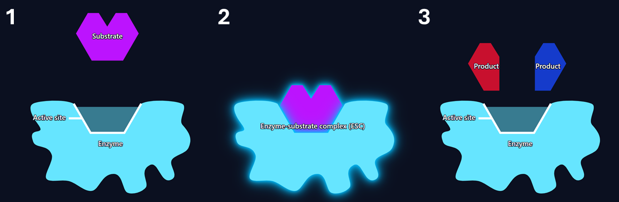
This is called the lock-and-key model because the enzyme’s active site is like a lock and the substrate like the key to that lock. Much like the way that only a key with the right shape can open a lock, only a substrate with a suitable shape can enter the active site, so we need thousands of different enzymes for the thousands of types of substrates. Also, a bit like how the master-key to your school building can open all of the different locks, some substrates can work with more than one enzyme, and some enzymes can work with more than one (similar) substrates. The scientific word to describe the relationship between enzyme and substrate is specificity: enzymes are specific to their substrates because they only work with specific substrates that are complementary to their active site.
Depending on the enzyme, this can take place anywhere between less than one to several hundred thousand times in a single second.
What happens when the active site changes shape?
The lock and key model shows how important the shape of the active site is. Sometimes an enzyme’s active site can change shape so that it is no longer complementary to the substrate. Small changes to the shape of the active site can make it harder for the substrate to enter, and make the substrate react more slowly once it does, resulting in reduced activity (rate) of the enzyme. Sometimes there are large changes to the active site and the wider 3D structure of the enzyme, which mean the substrate no longer fits at all and the enzyme no longer functions as a catalyst; at this point we say the enzyme has become denatured, the process of denaturation is often irreversible, especially when it results from high temperatures.
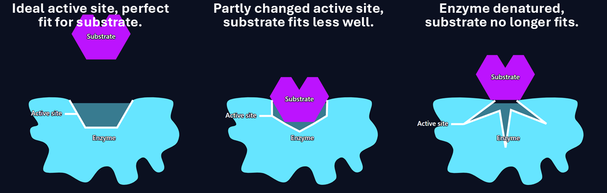
Enzymes can be denatured by the effects of both pH and temperature.
- Enzymes have a pH at which their active site is the ideal shape for the substrate and the enzyme works fastest, this is called the optimum pH. Small increases in pH above or below the optimum lead to small changes to the active site’s shape, so the enzyme still works but at lower activity (rate); large changes in pH above or below the optimum can denature the enzyme so the substrate no longer fits and the enzyme is no longer active. Note: If you are studying IB/A-Level biology, it is worth noting that pH also affects the protonation of the R-groups of the amino acids within the active site (this means whether they have gained or lost protons as a result of pH), influencing how well they bind with the substrate, so the effect of pH is a little more complex than just changing the active site.
- Enzymes have a temperature at which they work at their fastest rate, known as the optimum temperature. Small temperature increases above the optimum lead to small changes to the active site’s shape, so the enzyme still works but at lower activity (rate); large changes in temperature above the optimum can denature the enzyme so the substrate no longer fits, and the enzyme is no longer active. Temperature decreases below the optimum do not generally denature enzymes, and reduced activity is more to do with reduced particle movement, and thus collisions.
What is the induced fit model?
When first learning about how enzymes work, students are generally taught the lock-and-key model as it is an easy first step towards understanding, but it is limited because it makes enzymes seem like they have a fixed, rigid shape, when in reality their shapes undergo constant, small changes.
In the induced-fit model of enzymes, there is still an active site, and there is still a substrate, the difference is in how the active site changes during the process. Before a substrate docks with the enzyme, the shape of the active site is broadly complementary to the substrate; however, as the substrate docks the active site changes shape to become a better fit. Step-by-step, the induced fit model works something like this:
- The substrate enters the active site whose shape only approximates that of the substrate.
- As the substrate moves into the active site, interactions between the substrate and enzyme cause the active site to change shape, improving its fit to the substrate.
- An enzyme-substrate-complex The active site holds the substrate in the right position for a chemical reaction to happen which turns the substrate into the products.
- The products leave the active site, and the active site returns to its initial shape, ready to receive another substrate.
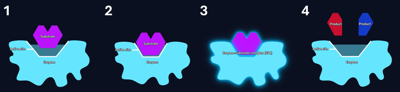
At post-16 studies, it is generally more appropriate to learn the induced fit model as it is a more accurate representation of how enzymes work. This is not to say that the lock-and-key model is entirely wrong, just that it contains reasonable simplifications and that the induced fit model is more complete because it removes one of these simplifications.
Try this in the simulation:
- With Enzyme model set to Lock-and-key, click Start to see the lock and key mechanism working, toggle Show labels to On if needed.
- Try adjusting the Active site distortion slider to 20%, notice that the active site changes shape and the enzymes works a little more slowly. Set it to 30% to see how an even greater change results in the enzyme working extremely slowly. Now try 40%, at this point, the enzyme is denatured and no longer functions.
- Toggle the Reaction type between Breakdown and Synthesis to see the difference between the two types of reaction.
- Toggle the Substrate count to Many and Reaction type to Breakdown and let it run for 30 seconds or so and note the reaction rate. Now set the Active site distortion slider to 20% and wait another 30 seconds, notice that the reaction rate is slower because there is a worse fit between the enzyme and substrate.
- Toggle the Enzyme model to Induced fit, notice now how the active site does not start out at quite the right shape, but changes as the substrate enters it.
Temperature
The temperature at which an enzyme shows the greatest activity (works fastest) is called the optimum temperature. The relationship between enzyme activity and temperature is determined by the balance of two factors:
- Particle collisions: At colder temperatures, molecules move more slowly, which means there are fewer collisions between enzymes and substrates resulting in reduced activity. As the temperature increases, the molecules move faster, which means there are more frequent collisions so the enzyme activity increases. At higher temperatures, molecules also collide with more energy, meaning they are more likely to have the activation energy for the reaction, and are less likely to simply bounce off each other.
- Active site shape: The lock-and-key model of enzymes tells us that functioning of enzymes depends on the active site having a complementary shape to the substrate. At temperatures above the optimum, the enzyme’s protein shapes can begin to unfold, changing the shape of the active site, reducing its complementarity and reducing its activity. At temperatures significantly above the optimum, the change to the active site can be so great that the substrate no longer fits, its activity drops to zero and the enzyme is described as denatured. Such denaturing is typically irreversible.
Enzymes work fastest at their optimum temperature because this is when the combination of high collision frequency and highly complementary active site shape allows enzyme-substrate complexes to be formed at their highest rate. The optimum temperature of the enzymes found in the human body is typically around 40 °C (although it varies significantly between enzymes), slightly above the human body temperature of 37 °C.
The graph of enzyme activity vs temperature for a given enzyme looks something like the below.
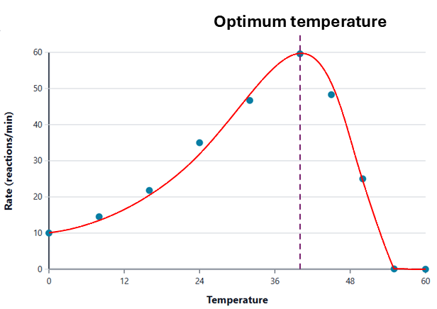
The graph slopes upwards with an increasing gradient towards the optimum temperature as the molecules move faster, which increases the frequency and energy of collisions, leading to a greater rate of enzyme-substrate complex formation. At the optimum temperature, collisions are happening at high frequency whilst the active site is still highly complementary. Beyond the optimum temperature, enzyme activity starts to fall because the increase in collisions is more than offset by changes in the shape of the active site, making it harder for substrates to bind to it. At significant temperatures above the optimum, the active site changes shape so much that the enzyme becomes denatured, and activity drops to zero.
Try this in the simulation:
- Set the Temperature slider to 0 °C, toggle Show countdown timer to on, leaving its slider at 30 s, and press the Start button and note the rate after 30 s. Repeat this with the temperature set to 40 °C. Notice how, whilst the shape of the active site stays the same, the rate increases because the particles are moving faster so they collide more frequently and with more energy.
- Repeat the above but with the temperature set to 60 °C; note how, despite the increase in particle movement speed, the rate drops to zero because the active site changes shape so much that the substrates can no longer bind to it.
- Repeat the above but with the Optimum temperature slider set to a different value. Notice how the general pattern of the results is the same, but their peak occurs at a different temperature.
- Toggle Experiment mode to On, then click the Experiment settings button. Choose a range of temperatures both above and below the optimum, then click the Confirm settings button. Press Start, and toggle Auto run if you want the sim to automatically complete all the runs you selected. Once complete, view the graph to see the shape of the relationship between temperature and enzyme activity.
pH
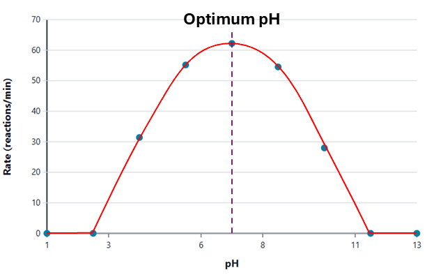
The graph below shows the relationship between pH and enzyme activity. As you can see, there is an optimum pH at which the enzyme works at the maximum rate. It then decreases in activity with small changes in pH above or below the optimum, with larger changes in pH resulting in larger decreases in activity until it eventually drops to zero. Most enzymes in the body have an optimum pH of around 7, although this can be as low as pH 1.5–2 for the digestive enzyme pepsin in our stomachs, and as high as pH 10 for enzymes such as alkaline phosphatase.
The shape of the graph can be explained by the effects of two factors:
- Active site shape: At the optimum pH, the enzyme’s active sites have the most complementary shape for the substrate, meaning that substrates readily bind with the enzymes to form enzyme-substrate complexes, leading to a high rate of reaction. As the pH changes away from the optimum, the active site begins to change shape, making it less complementary and making it harder for substrates to bind. This reduces the rate of enzyme-substrate complex formation and thus enzyme activity. The greater the change in pH, the greater the change to the active site and the more the enzyme’s activity drops. With small-to-medium deviations in pH away from the optimum, changes to the active site are generally reversible; at large changes in pH, the enzyme can become irreversibly denatured and its activity drops to zero.
- R-group protonation (post-16 only): Enzymes are made from proteins, which are themselves made from amino acids. Amino acids contain R-groups, the name given to a large number of different side chains that are unique to each amino acid. It is the interactions between R-groups that determine the folding of the enzyme’s protein chain and thus its 3D shape. Changes in pH can change the protonation of the R-groups, which means they can gain or lose H+ ions, changing the way they interact with other R-groups and thus the shape of the enzyme and its active site. Bigger changes in pH lead to bigger changes in protonation. Changes in protonation also affect the way the substrate binds with the enzyme, meaning it is not simply about the active site.
Try this in the simulation:
- With the pH slider set to 5 and Show countdown timer toggled to On, press Start, then note the rate once the timer stops. Repeat at pH 7 and then pH 9. Notice that the reaction is fastest at pH 7 and slower but similar at pH 5 and 9. Notice also that the only thing that changes is the shape of the active site: it is ideal at pH 7 and slightly changed at pH 5 and 9, while particle speed remains unchanged.
- Repeat a similar experiment but with the Optimum pH slider set to a different pH. You should see a similar pattern but with the optimum at a different pH.
- Toggle Experiment mode to On, then click the Experiment settings button. Choose a range of pH values both above and below the optimum, then click the Confirm settings button. Press Start, and toggle Auto run if you want the sim to automatically complete all the runs you selected. Once complete, view the graph to see the shape of the relationship between pH and enzyme activity.
Substrate concentration
The graph below shows the relationship between the concentration of substrate molecules and the rate of an enzyme-controlled reaction.
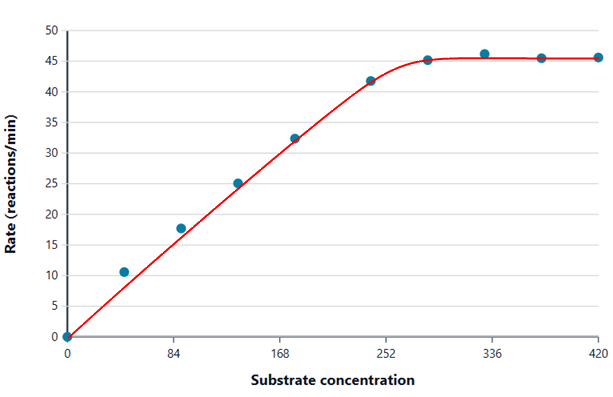
As the concentration of the substrate increases, the rate of that reaction also increases because there is an increased frequency of collisions between substrate and enzyme molecules, so there is an increased rate of enzyme-substrate complex formation. At high substrate concentrations, further concentration increases lead to smaller and smaller increases in the rate until a maximum rate is achieved. This happens because there is a limit to the number of times each second that a substrate can bind to the active site, react and the products vacate the active site. As an analogy: if I gave you one chocolate to eat each hour, the rate you ate chocolates would be one per hour; if I gave you one each minute, the rate you ate them would be 60 per hour, but if I gave you one each second the rate you eat them would not be 3,600 per hour because it takes you longer than a second to eat each one.
Note: In this sim, it consistently seems that at the highest substrate concentrations, the activity actually decreases slightly. This is not realistic behaviour and is a result of trying to model highly complex 3D systems in a simplified 2D form. The key thing to take away here is that enzyme activity increases with substrate concentration, but stops increasing any further once the substrate concentration has reached a certain level.
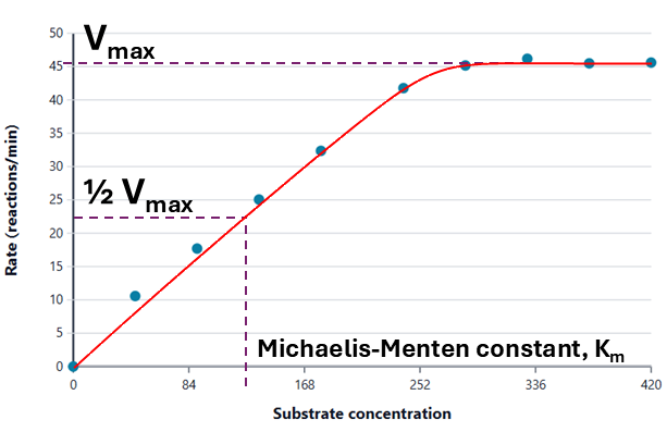
Post-16: The maximum rate at which an enzyme works is known as Vmax. Vmax varies widely depending on the enzyme and is fixed for a given enzyme, for a given temperature and enzyme concentration. The substrate concentration at which half of Vmax is reached for a particular enzyme is known as its Michaelis constant, Km. This is considered an indicator of how effectively an enzyme binds to its substrate, with lower values indicating stronger binding because the enzyme is able to achieve higher activity at lower concentrations.
Try this in the simulation:
- Set the Substrate concentration slider to 150, toggle Show countdown timer to On, and then click Start. Repeat this with the concentration set to 300 and you should find the rate roughly doubles. Now increase the concentration to 420 and repeat. You should see that the rate does not significantly change because the enzymes are already working at their maximum rate.
- Toggle Experiment mode to On, then click the Experiment settings button. Choose a range of substrate concentrations, then click the Confirm settings button. Press Start, and toggle Auto run if you want the sim to automatically complete all the runs you selected. Once complete, view the graph to see the shape of the relationship between substrate concentration and enzyme activity.
Competitive inhibition (post-16 only)
Some molecules can have a sufficiently similar shape to an enzyme’s substrate that they can also bind to the active site; this binding can be reversible or permanent. In situations like this, the substrate finds itself in competition for the active site and the molecule it is competing with is called a competitive inhibitor. By occupying the active sites for some of the time, competitive inhibitors reduce the rate of enzyme-substrate complex formation, and thus the overall level of enzyme activity.

In the image above, the left enzyme molecule has a purple substrate bound to its active site, showing the enzyme functioning normally; in the right one, an orange competitive inhibitor has bound to the active site, preventing a substrate molecule from binding and forming an enzyme-substrate complex.
As the concentration of competitive inhibitor molecules increases, the chance of substrates forming enzyme-substrate complexes decreases, so enzyme activity decreases, as can be seen in the graph below.
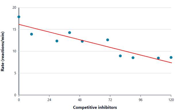
In the presence of a competitive inhibitor, enzyme activity is reduced below its maximum, Vmax. However, if you increase the substrate concentration sufficiently, it increases the likelihood that the molecule binding to the active site will be a substrate rather than an inhibitor, and enzyme activity will once again approach Vmax. Competitive inhibition does not change Vmax, but does change the effective Km because a higher substrate concentration is required to achieve half of Vmax.
Try this in the simulation:
- Set the Competitive inhibitors slider to 0, toggle Show countdown timer to On, and then click Start. Repeat this with the slider set to 40 and then again at 80. You should see the reaction rate drop by about 20% at 40 inhibitors and 40% at 80 inhibitors.
- Repeat the above but with the Inhibitor binding strength slider set to 60 s. This increases the average time that an inhibitor binds to the enzyme before being released. You should see that enzyme activity significantly decreases as the active sites are occupied by inhibitors for a greater proportion of the time, reducing their availability to form enzyme-substrate complexes.
- With the Competitive inhibitors slider set to 30, experiment with a range of different Substrates values, and note how the rate increases significantly with increasing substrate concentration as the substrates are able to compete more effectively with the inhibitors for access to the active sites.
- Toggle Experiment mode to On, then click the Experiment settings button. Choose a range of competitive inhibitor values, then click the Confirm settings button. Press Start, and toggle Auto run if you want the sim to automatically complete all the runs you selected. Once complete, view the graph to see the shape of the relationship between competitive inhibitor concentration and enzyme activity.
Non-competitive inhibition (post-16 only)
Some molecules are able to bind to regions on the enzyme other than the active site in ways that do not prevent the formation of enzyme-substrate complexes but prevent the enzyme from catalysing the reaction, causing the activity of inhibitor-bound enzymes to drop to zero.

In the image above, the left enzyme is unaffected by a non-competitive inhibitor, so it will be able to catalyse a reaction as normal. In the right one, a non-competitive inhibitor molecule has bound to the enzyme, so even though a substrate has also bound to the active site, no reaction can take place as a result of the inhibitor.
As the concentration of non-competitive inhibitor molecules increases, the proportion of enzyme molecules with inhibitors bound to them increases, so enzyme activity decreases because there are fewer unaffected enzyme molecules able to catalyse reactions.
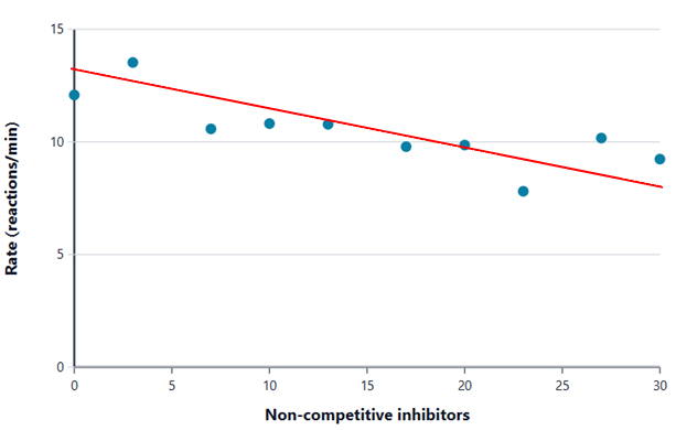
In the presence of a non-competitive inhibitor, enzyme activity is reduced, and increasing the substrate concentration will not return it towards Vmax, as the inhibitors are not competing with substrates for access to the active site and so will have the same inhibiting effect regardless of substrate concentration. As such, the presence of a non-competitive inhibitor reduces Vmax but does not affect Km. Removing the inhibitor will restore the enzyme to normal functioning. It is worth noting that the sim uses a pure non-competitive model but real inhibitor behaviour can be more complex than this.
Try this in the simulation:
- Set the Non-competitive inhibitors slider to 0, toggle Show countdown timer to On, and then click Start. Repeat this with the slider set to 30 and then again at 60. You should see the reaction rate progressively decrease as more enzymes spend time with inhibitors bound.
- Repeat the above but with the Inhibitor binding strength slider set to 60 s. This increases the average time that an inhibitor binds to the enzyme before being released. You should see that enzyme activity significantly decreases as the enzymes spend longer bound to inhibitors and unable to make reaction progress.
- Toggle Experiment mode to On, then click the Experiment settings button. Choose a range of non-competitive inhibitor values, then click the Confirm settings button. Press Start, and toggle Auto run if you want the sim to automatically complete all the runs you selected. Once complete, view the graph to see the shape of the relationship between non-competitive inhibitor concentration and enzyme activity.